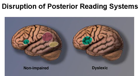
The continuing research into brain physiology and the connection to dyslexia is providing more answers in order to understand what dyslexia is, how we can diagnose it, how it is related to language and to reading, and the remediation that works.
Dyslexia is related to the susceptibility of some genes to develop differently during fetal development which is the biological condition of dyslexia. Children with dyslexia often show symptoms of other neurobiological conditions, including ADHD. The use of cutting edge technology, including fMRI’s and scans, helps identify the genetic markers of dyslexia and gives us the opportunity for earlier interventions.
Dyslexia is not related to intelligence or motivation. Fifteen to twenty percent of the population has a reading disability. Dyslexics are talented in the arts or in using their hands and are very creative. Dyslexia is language-based and refers to a cluster of difficulties in spelling, reading, writing and speaking. It is a life-long challenge and has a different impact at different times in one’s life. (Lyon, Shaywitz & Shaywitz, Overcoming Dyslexia)
The recent use of fMRI (functional magnetic resonance imaging), DCM (Dual Causal Modeling) and PET (positron emission tomography) scans in measuring brain activity in all aspects of reading and language, visual and auditory processing, phonological processing, orthographic responses and spelling, rapid automatic naming, memory and fine motor skills of dyslexics and non-dyslexics point to anatomical differences and rates of activation of processes in different parts of the brain.
The parts of the brain involved in conscious activities such as learning and thinking include the cerebrum and the cerebellum, where the major actions involved in balance, coordination and writing are activated, and the amygdala and hippocampus, in the medial lobe, related to emotion and memory.
Let’s talk about the parts of the cerebrum impacted in reading.
Frontal Lobe
responsible for speech, executive function, reasoning, planning, problem solving, behavior, regulating emotions and consciousness
Parietal Lobe
controls sensory perceptions, links spoken and written language to memory, gives meaning to what we hear and read, affects math and spelling
Occipital Lobe
controls sensory perceptions, visual perceptions and identification of letters
Temporal Lobe
involved in verbal memory and understanding of language
LEFT Parietal-temporal Area
involved in word analysis and decoding as well as mapping letters and words into corresponding sounds
LEFT Occipital-temporal Area
involved in automatic rapid access to word analysis and fluent reading
Gray Matter
involved with phonological awareness
White Matter
helps the nerves transfer information so that the brain regions can communicate effectively, needed for reading
Corpus Callosum
the communication bridge between the cerebral hemispheres
The Brain without Dyslexia
- Good readers show more activation in the left hemisphere in ALL areas needed for reading: phonological processing, orthographic mapping for sound/letter connections for spelling and writing, interpretation of sounds and faster activation of the brain for rapid automatic naming responses.
- They show less activity in the right hemisphere.
- There are metabolic differences in blood flow and physical differences in size.
The Brain with Dyslexia
- Dyslexics show disruptions in the rear reading system in the left hemisphere, critical for reading fluently.
- There is more activation in the less efficient right hemisphere, thought to be a compensation method.
- There is a different distribution of metabolic activation when working on the same tasks as non-dyslexics.
- There is greater activation in the lower frontal area.
- Less activity in the Left Parietal-temporal lobe required for phonological processing is seen where identifying and manipulating individual sounds and the structure of words.
- There is less activity in the Left Occipital-temporal lobe that affects the “orthographic” mapping or understanding of letters into sounds, auditory processing and interpretation of sounds, difficulty with rapid rate of information coming in or phonology of sounds.
- There is also less gray matter to help with transfer of information of language (phonological processing).
- Less white matter disrupts communication of information.
- There are anomalies in the size of the corpus callosum.
- There is less memory storage capacity for phonological coding or naming.
The good news derived from all the neuroscience studies is that they can now be applied to the classroom to help students perform better. With proper identification, remediation can begin sooner. There are “brain-friendly” teaching methodologies, schools can implement academic modifications and with the availability of assistive technology, the differentiated needs of the dyslexic learners are being met.
In summary, educational neuroscience offers methods for identifying early markers for recognizing those at risk for reading. Analyzing brain imaging of students with reading disabilities shows us what the brain looks like when there are no interventions and when interventions are put into place providing specific remedial programs. What we see is increased activation in the left hemisphere, important for reading. After one year of intervention, we see increased activity in the occipito-temporal region and decreased activity in the right hemisphere important for automatic, fluent reading.
Sharing knowledge about brain functioning is one way of demystifying dyslexia and helps with explaining how the brain functions when language processing is concerned. It is very important for both parents and teachers to truly understand what dyslexia is and is not. Once the misconceptions are cleared up, and explicit instruction is provided, we can begin to see examples of “neuroplasticity” and changes in brain physiology.
Submitted by Cecile Selwyn, M.Ed Ed.S
Director of Commonwealth Learning Center, Needham
Header Image: Sally Shaywitz, Overcoming Dyslexia, 2003 from The Yale Center for Dyslexia and Creativity, dyslexia.yale.edu
(1) Brain Lobes Functions image from Headway, the brain injury association, www.headway.org.uk
(2) At Risk Reader image from The Morris Center, Picture of Dyslexia PowerPoint, themorriscenter.com


Jaydin Skinner says: